
New release: Altered muscle development in Beckwith-Wiedemann syndrome macroglossia
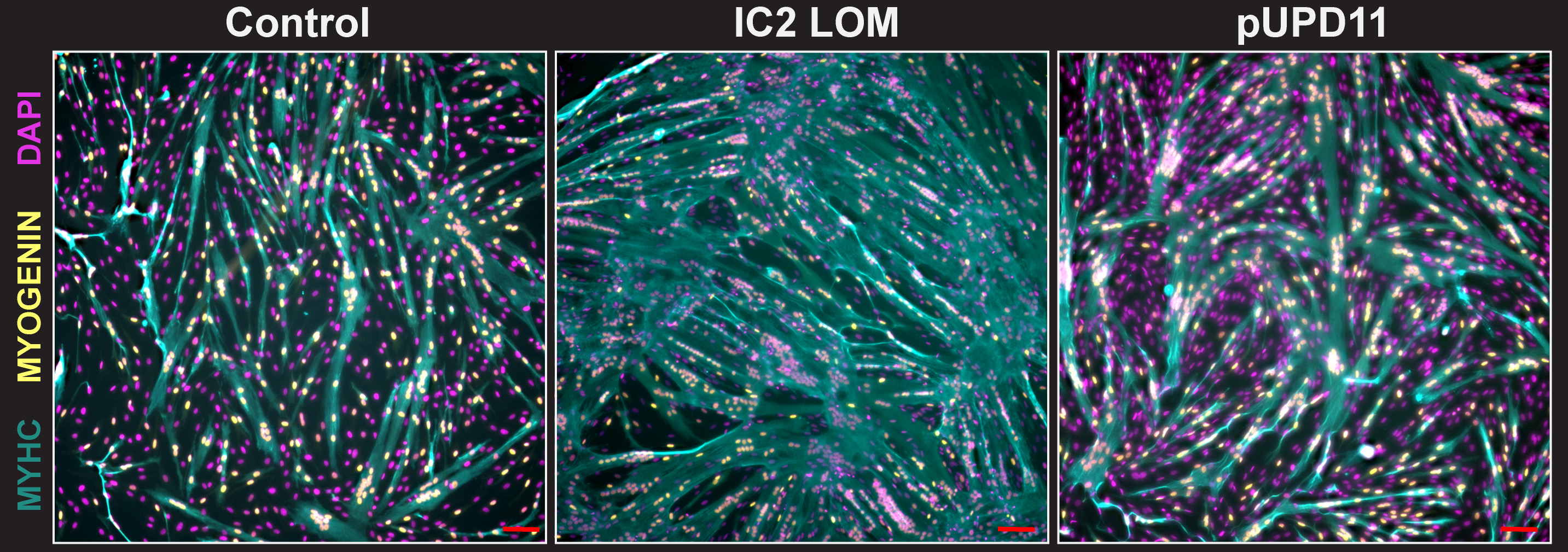
September 15th, 2026FaceBase has released a new dataset from the Kalish Lab at Children’s Hospital of Philadelphia (CHOP) containing bulk RNA sequencing data from muscle stem cells (satellite cells) isolated from the tongues of children with and without Beckwith-Wiedemann syndrome (BWS). BWS is an overgrowth disorder caused by genetic and epigenetic changes on chromosome 11p15, and an enlarged tongue (macroglossia) is one of its most common features. By comparing cells from two molecular subtypes of BWS with nonsyndromic controls as they develop into skeletal muscle, these data can help researchers identify the molecular pathways behind altered tongue muscle development in BWS macroglossia.
Contributors: Elisia D. Tichy, Jennifer M. Kalish (Children’s Hospital of Philadelphia)
Description:
This dataset contains very low input bulk RNA-seq data from tongue-derived satellite cells. Human pediatric samples include nonsyndromic controls and two BWS molecular subtypes: IC2 LOM (loss of methylation at imprinting control region 2) and pUPD11 (paternal uniparental disomy of chromosome 11). After FACS isolation of satellite cells (defined as CD45-/CD324-/CD56+/CD82+), cells were plated in myoblast growth media for 6 days and then switched to differentiation media for 2 days.
Libraries were prepared from 5 ng of total RNA and sequenced paired-end on an Illumina NovaSeq X. The dataset includes FASTQ files (4 per sample) for 3 controls, 3 BWS IC2 LOM, and 3 BWS pUPD11 samples.
Supported by the National Institute of Dental and Craniofacial Research (award R01DE033646).
Accessing the data
This is restricted-access human data (data use limitation: General Research Use). To gain access, you must first complete the process outlined here.
Explore the dataset in the FaceBase Data Browser
FaceBase Dataset:
Elisia D Tichy, Jennifer M Kalish. Gene expression patterns in pediatric tongue-derived satellite cells during differentiation (Bulk RNAseq). FaceBase Consortium https://doi.org/10.25550/A5-VYP4 (2026).
Associated Publication:
Elisia D Tichy, Anna T Nguyen, Mariah A Byrne, Rose D Pradieu, Gavriela Kalish-Schur, Mara Fallon, Snehal Nirgude, Darryl Kinnear, Harry P Kozakewich, Jennifer M Kalish. Myogenic dysregulation underlies human tongue overgrowth in Beckwith-Wiedemann syndrome. iScience 29(10), 117575 https://doi.org/10.1016/j.isci.2026.117575 (2026).
New release: KDM6B Acts via BMP Signaling During Root Morphogenesis
September 8th, 2026FaceBase has released a new dataset from the Chai Lab at USC examining how the tissue surrounding a developing tooth root recruits the sensory nerves that will eventually supply it.
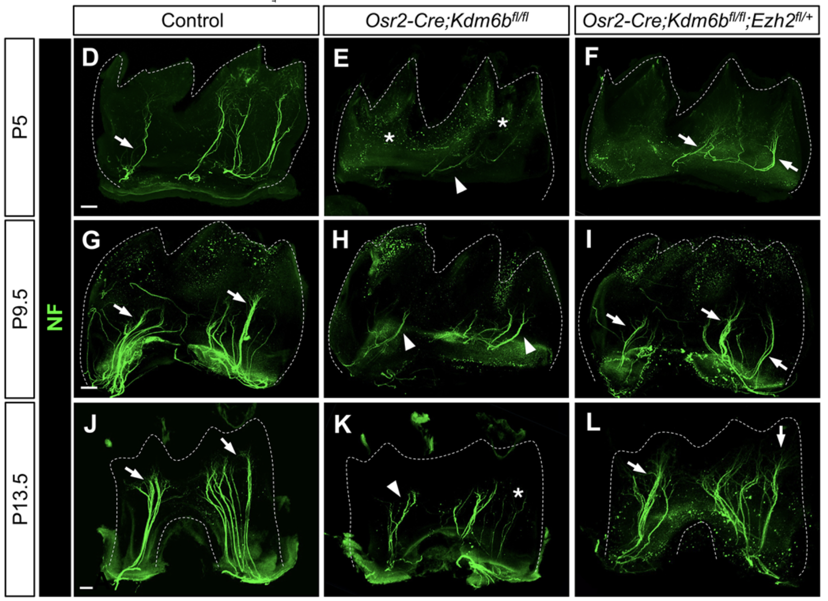
Developing organs do not simply receive innervation; they have to instruct it. Tooth root development is a useful place to watch that happen, because root morphogenesis and sensory innervation begin and progress at the same time in the same tissue. What has been unclear is which signals the surrounding mesenchyme uses to direct incoming axons, and how those signals are controlled.
Contributors: Heliya Ziaei, Mingyi Zhang, Tingwei Guo, Jifan Feng, Lin Meng, Angelita Araujo-Villalba, Junjun Jing, Thach-Vu Ho, Yang Chai (USC)
Description:
Apical molar mesenchyme was collected at postnatal day 3.5 from control (Kdm6bfl/fl), mutant (Osr2-Cre;Kdm6bfl/fl), and rescue (Osr2-Cre;Kdm6bfl/fl;Ezh2fl/+) mice and profiled two ways. Bulk RNA-seq (n=3 biological replicates per genotype, each pooled from four animals) captures the transcriptional consequences of losing Kdm6b in dental mesenchyme. CUT&RUN sequencing for H3K27me3, with matched IgG controls, maps the repressive chromatin marks that mediate them. All libraries were sequenced on an Illumina NovaSeq.
The associated study, published in PNAS in 2026, found that mesenchymal loss of Kdm6b reduces expression of the BMP antagonist Bambi through changes in H3K27me3, which overactivates BMP signaling and in turn suppresses the neurotrophic factor NGF. Without that mesenchymal support, trigeminal axons fail to enter and branch into the dental pulp, and root development is disrupted. Removing one copy of Ezh2, which opposes Kdm6b, partially restores Ngf expression, innervation, and root formation.
Supported by the National Institute of Dental and Craniofacial Research (award R01DE012711).
Accessing the data
This dataset is openly available. Files can be downloaded directly from the dataset record; no application is required.
FaceBase Dataset:
Heliya Ziaei, Mingyi Zhang, Tingwei Guo, Jifan Feng, Lin Meng, Angelita Araujo-Villalba, Junjun Jing, Thach-Vu Ho, Yang Chai. KDM6B Acts via BMP Signaling During Root Morphogenesis. FaceBase Consortium https://doi.org/10.25550/A2-0AAR (2026).
Associated Publication:
Ziaei H, Zhang M, Guo T, Feng J, Meng L, Araujo-Villalba A, Jing J, Ho TV, Chai Y. Epigenetic regulation of mesenchymal BMP signaling directs postnatal organ innervation. Proc Natl Acad Sci U S A. 2026 Jul 7;123(27):e2605881123. doi: 10.1073/pnas.2605881123. Epub 2026 Jun 30. PMID: 42378299; PMCID: PMC13342922.
New release: Eyelid Kinematics and Orbicularis Oculi Electromyography
September 1st, 2026FaceBase has released a new dataset from the Clites Lab at UCLA that captures how the human eyelid moves and which parts of the muscle drive that motion during five distinct eyelid behaviors. It is our first dataset supported by the National Eye Institute.
Every blink spreads tear film across the cornea and clears debris from the ocular surface. Facial paralysis takes away the ability to blink, which can cause damage to the eye or infection. Efforts to restore blinking through electrical stimulation have been limited by a fundamental gap: no one has measured, at high resolution, what drives natural blink at the muscle level.
Contributors: Tyler Clites, Jinyoung Kim, Daniel Rootman (UCLA)
Description:
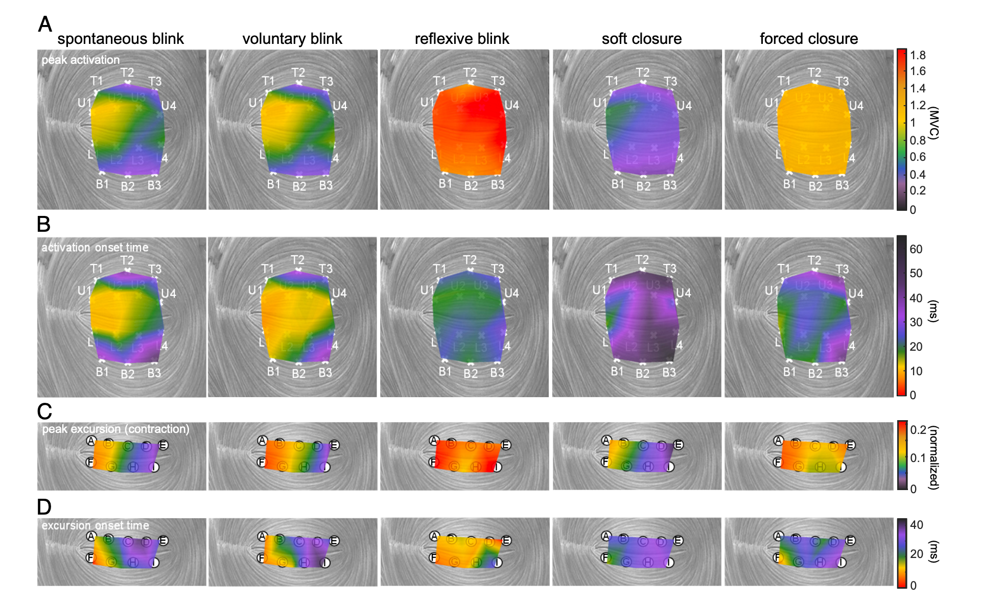
Eight adults without eyelid paralysis or other eyelid pathology (ages 22-33) performed spontaneous blink, voluntary blink, reflexive blink, soft closure, and forced closure while two systems recorded simultaneously: three-dimensional motion capture of markers along the upper and lower eyelid margins at 400 frames per second, and distributed intramuscular EMG from 14 bipolar fine-wire electrode pairs within the orbicularis oculi. The streams were hardware-time-synced, so muscle activation can be aligned frame-by-frame with the resulting eyelid motion.
The associated study, published in PNAS in 2025, found that different eyelid behaviors are produced by distinct patterns of activation across muscle segments, providing a clue as to why previous stimulation methods have been ineffective. Activating the whole eyelid at once tends to elicit a protective reflexive blink rather than the natural blink that keeps the eye wet and clear.
Supported by the National Eye Institute (award 1R21EY036680).
Accessing the data
This is restricted-access human subjects data. Researchers must complete the process described in the FaceBase data guidelines before access can be granted. Data use is limited to General Research Use (GRU) and Not-for-profit Use Only (NPU).
FaceBase Dataset:
Tyler Clites, Jinyoung Kim, Daniel Rootman. Eyelid Kinematics and Orbicularis Oculi Electromyography - Persons Without Facial Pathology. FaceBase Consortium https://doi.org/10.25550/88-28BJ (2026).
Associated Publication:
Kim J, Shirriff A, Cornwell JN, Mutis MPQ, Delis E, Wang S, Rootman DB, Clites TR. Human eyelid behavior is driven by segmental neural control of the orbicularis oculi. Proc Natl Acad Sci U S A. 2025 Aug 12;122(32):e2508058122. doi: 10.1073/pnas.2508058122. Epub 2025 Aug 7. PMID: 40773233; PMCID: PMC12358864.
New release: Sensory nerve-derived signaling coordinates oropharyngeal structural organization that supports suckling and vocalization in neonatal mice
August 25th, 2026A new dataset from the Chai lab at the University of Southern California is now available in FaceBase, combining single-cell RNA sequencing, bulk RNA sequencing, and SeqFISH spatial transcriptomics to examine how sensory nerves shape the developing soft palate.
Contributors: Sa Cha, Jifan Feng, Tingwei Guo, Lin Meng, Peng Chen, Calista Ly, Thach-Vu Ho (Center for Craniofacial Molecular Biology, Herman Ostrow School of Dentistry, University of Southern California); Pedro A. Sanchez-Lara (Section of Medical Genetics, Department of Pediatrics, Guerin Children’s at Cedars-Sinai Medical Center); Lauren E. McElvain and Jeffrey D. Moore (Department of Biological Sciences, Dornsife College of Letters, Arts and Sciences, University of Southern California); Yang Chai (Center for Craniofacial Molecular Biology, Herman Ostrow School of Dentistry, University of Southern California)
Description:
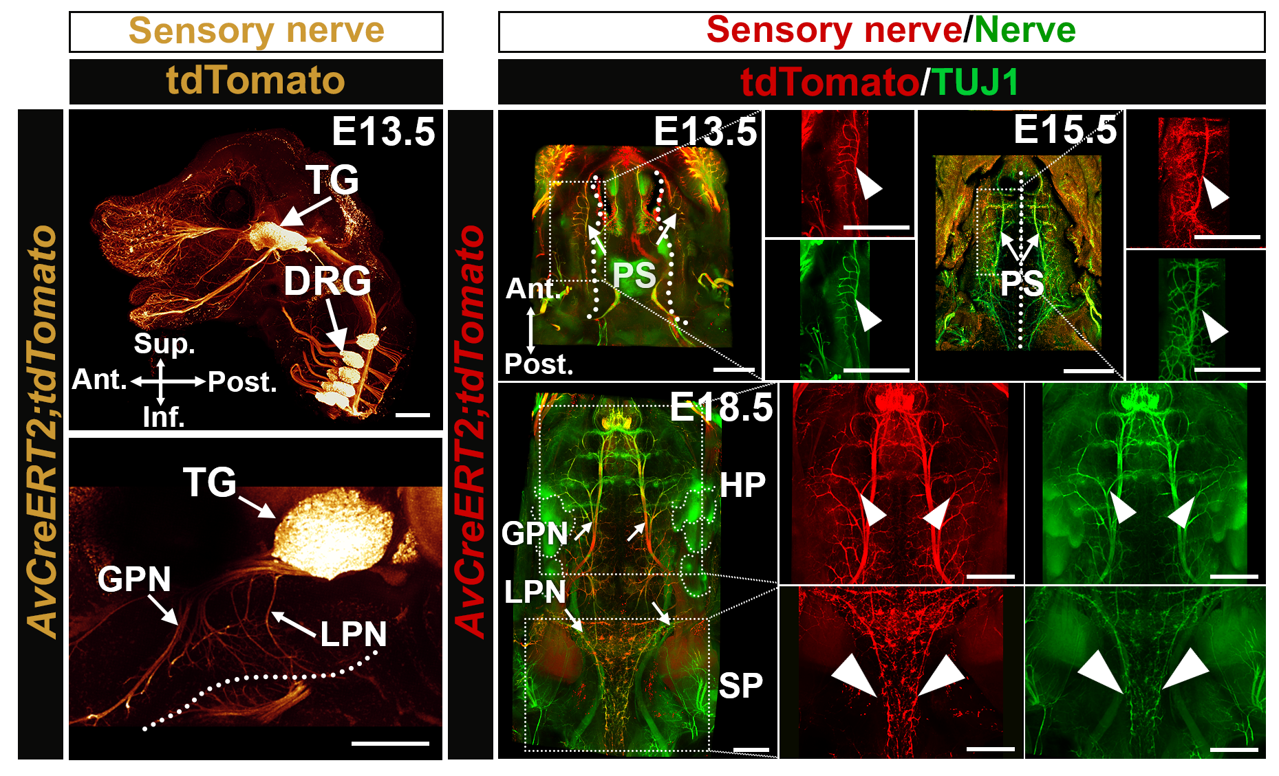
Proper oropharyngeal function is essential for suckling, feeding, and speech, and relies on the coordinated development of the palate, oropharyngeal musculature, and the cranial nerves that control them. Disruption of this integration leads to severe neonatal complications. However, how neuromuscular architecture is developmentally coordinated to support oropharyngeal function remains unclear. Using single-cell and spatial transcriptomics, we identify trigeminal nerve-derived GDF11 as a crucial regulator of soft palatal muscle architecture that acts through cranial neural crest-derived perimysial cells and is mediated by Akt-FoxO1-Thbs3 signaling to establish muscle structural integrity and bilateral continuity. To assess its functional relevance in vivo, we employ a battery of physiological assays to evaluate oropharyngeal function in neonatal mice and find that sensory neuron-specific Gdf11 deletion recapitulates soft palatal deformities and associated oropharyngeal dysfunction observed in individuals carrying GDF11 mutations, including impaired suckling, reduced oropharyngeal motor efficacy, and abnormal vocalizations. Pharmacological activation of AKT partially restores soft palatal muscle organization and ameliorates associated physiological deficits in Gdf11 mutant mice. Collectively, these findings demonstrate that sensory nerve-derived trophic signaling is indispensable for coordinated oropharyngeal morphogenesis and function, and establish a pre-clinical framework for the therapeutic approach targeting neuromuscular integration in congenital oropharyngeal disorders.
What’s in the dataset
The deposited data are embryonic. Samples come from trigeminal ganglion and palatal shelf tissue at E13.5 and E14.5, spanning eight experiments, 19 biosamples, and more than 25 files. Three assay types are represented:
- Single-cell RNA sequencing of dissected tissue
- Bulk RNA sequencing
- SeqFISH spatial transcriptomics, preserving positional information across the palatal shelves
Processing used Cell Ranger 9.0.0 and SGNlite, with sequencing on Illumina NextSeq 500 and NovaSeq platforms.
The data are open access and can be downloaded directly from FaceBase.
This work was supported by NIDCR grants R01 DE012711 and U01 DE028729 to Yang Chai. The U01 is a FaceBase data generation project, making this a case of FaceBase-funded research depositing its underlying data back into the repository.
FaceBase Dataset:
Sa Cha, Jifan Feng, Tingwei Guo, Lin Meng, Peng Chen, Calista Ly, Thach-Vu Ho, Pedro Sanchez, Lauren E. McElvain, Jeffrey D. Moore, Yang Chai. Investigating Oropharyngeal Development in Neonatal Mice Using scRNA sequencing, RNA sequencing, and SeqFISH. FaceBase Consortium https://doi.org/10.25550/8N-FGKC (2026).
Associated Publication:
Cha S, Feng J, Guo T, Meng L, Chen P, Ly C, Ho TV, Sanchez-Lara PA, McElvain LE, Moore JD, Chai Y. Sensory nerve-derived signaling coordinates oropharyngeal structural organization that supports suckling and vocalization in neonatal mice. Nat Commun. 2026 Jul 14. Advance online publication. https://doi.org/10.1038/s41467-026-74959-0
New release: Center for TMD IMPACT (C-TMD IMPACT) Patient and Clinician Needs Assessment
August 18th, 2026Two new datasets from the Center for TMD IMPACT (C-TMD IMPACT) are now available in FaceBase. Together, they offer a view of common needs and common barriers from both sides of the clinical encounter, and a starting point for researchers and clinicians looking to collaborate on addressing TMDs.
Contributors: Yang Chai, Jian-Fu Chen, and VyVy Nguyen (University of Southern California)
Description:
During the TMD IMPACT Collaborative R34 planning phase, the Center for TMD IMPACT at the University of Southern California conducted a needs assessment to better understand the needs of patients who experience symptoms of TMD and clinicians who treat them. Both surveys were administered in 2024 and the resulting data have been de-identified for open release.
Patient Needs Assessment
Center for TMD IMPACT Patient Needs Assessment includes survey responses from 103 patients seen at the USC Orofacial Pain and Oral Medicine Center between May and July 2024. The survey covered patient demographics, descriptions of symptoms, and descriptions of treatments. Patients identified six priority areas for improving TMD treatment: better coverage of treatment, faster and more immediate access to care, more frequent appointments, better information about TMD, more knowledgeable providers, and better bedside manner from clinicians.
Clinician Needs Assessment
Center for TMD IMPACT Clinician Needs Assessment includes survey responses from 17 practicing clinicians in the Los Angeles and San Francisco Bay areas who treat patients with TMDs, collected between June and August 2024. The survey covered clinician demographics, opinions on the current standard of treatment, and ideas for improving it. Clinicians pointed to three priority areas: addressing gaps in phenotyping and pain research, improving patient access to care, and improving quality of care through interdisciplinary practice, better educational curriculum and opportunities, and better technology and software.
Each dataset includes the survey response data, a data dictionary, and a PDF of the survey instrument. Both are open access under General Research Use (GRU) limitations and can be downloaded directly from FaceBase.
FaceBase Datasets:
Yang Chai, Jian-Fu Chen, VyVy Nguyen. Center for TMD IMPACT Patient Needs Assessment. FaceBase Consortium https://doi.org/10.25550/9N-634P (2026).
Yang Chai, Jian-Fu Chen, VyVy Nguyen. Center for TMD IMPACT Clinician Needs Assessment. FaceBase Consortium https://doi.org/10.25550/9N-634W (2026).